Here you can find the latest news and surgical articles.
19 June 2026
Machine Learning Predicts Fat Transfer Survival Rates and Clinical Implications for Cosmetic Procedures
Key Takeaways
Machine learning predicts individualized fat graft survival using patient, procedural, and postoperative data to estimate retention rates and guide surgical planning.
Gather standardized inputs from patient BMI, adipose tissue quality, harvest and processing methods, and follow-up retention to enhance model accuracy.
Feature selection prioritized adipocyte viability, graft volume, and fat processing technique while eliminating redundant variables to minimize overfitting and increase prediction accuracy.
Validate models with cross-validation and external datasets. Use clear performance metrics. Update models regularly to maintain generalization across populations and techniques.
Use these digital twin simulations to test techniques and personalize graft size, injection sites, and postoperative care. This reduces variability and supports evidence-based decisions.
Tackle existing challenges by developing multicenter registries, incorporating diverse datasets, creating clinician-accessible tools, and pursuing stepwise clinical implementation with ethical and regulatory review.
Machine learning predicting fat transfer survival rates applies machine learning algorithms to predict the survival rate of fat transfers. Models based on patient data, graft handling, and surgical factors provide a likelihood for volume retention.
Research shows that better planning and tailored care when forecasts inform choices. Clinicians can compare approaches and set realistic expectations with measurable retention forecasts.
The core briefly reviews approaches, datasets, and clinical impact for practice.
How AI Predicts
Machine learning models parse numerous variables simultaneously to predict fat graft survival. Models take patient data, surgical details and outcomes and detect patterns that humans can overlook. Here’s a quick map of how inputs turn into usable predictions for surgeons and patients.
1. Data Input
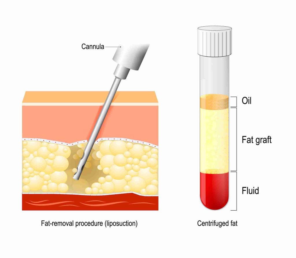
Collect patient demographics, body mass index, comorbidities, and prior procedures. Include adipose tissue traits like adipocyte density, cell size, and vascularity measured from imaging or biopsy. Record harvest details: liposuction method, cannula size, suction pressure, and volume aspirated.
Note processing choices such as centrifugation speed, filtration, washing, or enzymatic treatment and the time between harvest and grafting. Include graft site specifics: recipient bed type, vascular supply, and prior scarring.
Postoperative outcomes must feed back: measured retention percentage, fat necrosis incidence, inflammation markers, and any revisions. Standardize formats, with units in metric and defined times for follow-up in weeks or months, so data from clinics worldwide can combine.
For example, one study used large surgical registries to build models predicting blood loss with 94% accuracy. Similar scale and standardization help fat graft models.
2. Feature Selection
Pick predictors that matter: adipocyte viability assays, cell diameter ranges, and fat distribution (subcutaneous vs visceral). Add importance to graft volume, donor site, and processing method as these frequently alter survival.
Eliminate redundant lab values or duplicate imaging measures in order to keep models lean and avoid overfit. Run statistical tests and simple models to rank features. A variable that barely raises accuracy can be dropped.
Imaging-derived measures, such as CT-based fat metrics, could enhance predictions in the same way AI-derived pericardial fat did for CV risk. Leverage those imaging features when accessible, but construct fallback models using clinical information for low-resource environments.
3. Algorithm Training
Train supervised models on past examples mapping inputs to measured retention and complications. Use regression, tree ensembles, and neural nets depending on dataset size. Tune hyperparameters to find the best fit without overreach.
Confirm with cross-validation and separate holdout sets. Use data from multiple techniques to ensure wide applicability. External validation matters: test on other clinics' data as done in studies predicting 30-day readmissions or cardiovascular events beyond calcium scores.
That step exposes real-world lacunas and enhances safety.
4. Survival Prediction
Generate patient-specific survival estimates and retention curves. Expected retention percent, probability of necrosis, and confidence intervals. Display results graphically with charts or tables so surgeons can discuss trade-offs and strategize overcorrection or staged grafting.
Give probability scores to facilitate informed consent and risk stratification.
5. Performance Validation
Compare predicted retention with outcomes mean absolute error and ROC analysis. Note ranges in blood loss studies: the maximum error was approximately 188 mL and the minimum was approximately 0.22 mL, which helps set expectations for acceptable error in fat models.
Keep models current with new data and methods.
Critical Predictive Factors
About critical predictive factors. The key to predicting fat transfer survival is gaining a clearer picture of which variables are really significant. Below are the three general domains: patient, procedural, and postoperative data points, and the relevant factors within each that machine learning models should emphasize. The goal is practical: improve model accuracy and guide clinical choices that lead to stable graft outcomes.
Patient Data
Age is a core predictor. Older patients often show lower short-term survival and slower integration of grafted adipocytes. Include age as a continuous variable rather than bins to preserve signal. Record gender, average body mass index (BMI) in kg/m2, and baseline body fat percentage through bioimpedance or DEXA for finer resolution.
Note comorbidities. Obesity and diabetes impair microvascular health and raise resorption risk. Prior surgeries in the donor or recipient zones alter tissue planes and vascular beds, so include surgical history. Measure visceral and subcutaneous adiposity separately. Visceral fat relates to systemic inflammation.
Subcutaneous fat is more likely to provide viable graft tissue. Preoperative CT or ultrasound measures provide additional accuracy. Monitor perioperative weight change and lifestyle factors, such as smoking, alcohol, and exercise, that shift metabolic state and can alter graft survival over weeks to months.
Add consciousness level, acid–base balance (pH), AST, myoglobin, and need for mechanical ventilation when patients are medically complex. These six factors have demonstrated robust predictive value for 28-day survival in comparable surgical databases. For models for short-term predictions, go ahead and incorporate these lab and clinical data.
Procedural Data
Capture the exact harvest technique: tumescent liposuction, low-pressure suction, or power-assisted methods. Remember negative pressure values when possible. Higher pressure generally tends to damage adipocytes. Log processing steps: centrifugation speed and duration, filtration pore size, and whether enrichment (stromal vascular fraction or platelet-rich plasma) was used.
These decisions transform cell density and live cells. Document graft volume and injection technique, linear threading versus bolus, cannula size, layering plan. Annotate anatomical site exactly. The recipient bed’s vascularity is a powerful survival modifier.
Include intraoperative parameters such as graft droplet size distribution and tissue adherence observations. These intraoperative factors powered models that predict early revascularization and long-term retention.
Postoperative Data
Monitor volume retention at set intervals: 1 week, 1 month, 3 months, 6 months, and 12 months. Take consistent imaging or standardized photos and volumetric measurements. Document complications such as fat necrosis, fat embolism, infection, or graft failure with timestamps.
Evaluate cosmetic result, scar formation, and soft tissue regeneration on validated scales. For short-term outcome modeling, use metrics shown to work in related survival models: ROC curve analysis demonstrated strong discrimination with an area under the curve of 0.851 and decision curve analysis showed net benefit across threshold probabilities of roughly 20 to 80.
Sensitivity at a chosen threshold was about 0.704 with a 95% confidence interval of 0.714 to 0.850. It’s the long-term tracking of stability and longevity that closes the loop between prediction and real-world results.
AI Versus Tradition
Machine learning offers a different path from long-used clinical judgment and rule-based scores for estimating fat graft survival. Traditional methods rely on surgeon experience, gross intraoperative assessment, and simple clinical factors such as patient age, body mass index, smoking status, and comorbidities. These methods are quick and familiar, but they vary by operator and lack quantitative precision.
Rule-based risk scores and general risk equations in medicine, like the American Heart Association’s PREVENT equation for cardiovascular risk, show how fixed-factor models work. They combine age, sex, blood pressure, cholesterol, and diabetes to give a population-level estimate. That approach is useful, but it can miss nuanced, image-derived signals that affect fat graft take.
Machine learning models utilizing patient, imaging, and procedural variables predict graft survival more consistently. Algorithms can learn from massive data to weigh subtle patterns humans can’t easily measure. As a case in point, automated extraction of pericardial fat from imaging demonstrated how AI can provide value in cardiology by enhancing risk prediction when combined with traditional scores.
Like fat grafting, AI can quantify graft volume, surrogates of tissue perfusion on imaging, and donor-site characteristics, then integrate them with clinical data to optimize outcome predictions. Studies across medicine reveal event rates of close to 10% for some outcomes, highlighting the importance of more accurate prediction tools. In fat grafting, even small absolute improvements in prediction can change management for numerous patients.
In head-to-head comparisons, AI wins for both accuracy and reproducibility. Machine learning minimizes inter-operator variance and can generate probabilistic outputs instead of binary calls. That helps surgeons advise patients on realistic expectations and plan staged surgeries.
AI scales: many patients already receive imaging studies, and algorithms can extract relevant metrics without extra scans, making the approach practical and cost-conscious. In cardiology, AI-based imaging metrics have enhanced risk stratification particularly for patients at intermediate risk. Similar benefits are probable for borderline rejection cases for which conventional techniques yield ambiguity.
AI backs decisions with evidence by providing objective, data-informed risk projections in addition to conventional evaluations. Surgeons can utilize aggregate outputs, a clinician score and an AI likelihood, to opt for adjuncts like platelet-rich plasma, stem cell enrichment, or conservative overcorrection. Based on the information, this combination of AI and traditional methods provides a personalized view of risk, which could improve outcomes and reduce unnecessary reoperations.
Limitations remain: model bias, need for large diverse datasets, and clear validation across centers. We need continued trials and common registries to earn trust and demonstrate real-world value.
The Surgeon's Digital Twin
Think of a digital twin as a virtual replica of a patient that can run multiple ‘what if’ scenarios before a scalpel ever touches tissue. In fat transfer, these models integrate imaging, patient history, tissue metrics and machine learning to predict graft survival, distribution, and even complications like blood loss.
Digital twins have shown early promise across healthcare tasks from predicting blood loss in a 721 patient liposuction study to modeling brain tissue loss. Systematic reviews find they are about 80% effective at achieving their intended result, though the field is still nascent and needs further validation.
Simulating Technique
Modeling starts with data: high-resolution imaging, fat quality measures, vascular maps, and procedure variables feed into the twin. Machine learning then associates particular extraction, processing, and injection techniques with past survival rates.
This allows the twin to exhibit potential retention for a specific method. The twin predicts how altering graft volume or fat spreading will change ultimate retention. It can run dozens of volume distribution permutations in minutes and rank outcomes by predicted percent survival, contour smoothness, and complication risk.
Vascularization and local tissue quality are modeled as key survival determinants. This twin can simulate bad perfusion zones or scarred tissue and demonstrate how these areas will influence cell survival over days to months.
Steps to simulate fat grafting outcomes:
Take multimodal patient data (imaging, labs, history) and preprocess into a baseline format. Check data quality and normalize spatial coordinates for alignment.
Choose technique parameters, such as harvest method, centrifuge versus filtration, injection cannula size, and plane of injection, and encode them as model inputs.
Forward simulate with trained ML models to predict immediate cell survival, early ischemia effects, and long term retention. Include stochastic runs to demonstrate variability.
Calculate outcome metrics, such as percent retention, contour deviation, and risk scores, and display results as overlays on patient anatomy for surgeon review.
Personalizing Plans
Digital twins personalize operations for every patient with predictive outputs. They recommend graft size and location that satisfy aesthetic objectives while minimizing risk.
AI suggestions can direct decisions about fat manipulation, suggesting if a gentle wash or spin is likely better based on cell brittleness statistics. Injection site maps are confidence labeled.
Postop plans are personalized: recommended compression timing, activity limits, and follow-up imaging windows to catch early graft loss. These actions are designed to increase viable graft percentages and patient satisfaction.
Benefits of personalized plans:
Better match between patient anatomy and graft strategy
Reduced over‑ or under‑correction through precise volume forecasts
Customized processing and handling to protect cell viability
Targeted postoperative care to support graft integration
Reducing Variability
AI assistance standardizes critical steps so results are less reliant on individual skill. Data-driven checklists and visual overlays help to eliminate guesswork during surgery.
Practice across teams is more reproducible if everyone uses the same model outputs and decision thresholds. This helps enable uniform graft survival rates across clinics and geographies.
Checklist to reduce variability:
Ensure input data quality and standardized imaging protocols.
Use model‑recommended harvest and processing settings.
Follow injection maps with marked planes and volumes.
Apply prescribed postoperative regimen and monitoring schedule.
Current Limitations
ML predicting fat transfer survival rates is impeded by several practical and scientific limitations that must be transparent before clinical application. These limitations range from data quality to model generalizability across populations to workflow integration to deficiencies in foundational clinical research. The next subsections decompose these challenges and provide actionable tips to tackle them.
Data Scarcity
Limited, inconsistent datasets are a fundamental issue. Comprehensive, annotated datasets connecting patient factors, graft preparation, injection technique, imaging, and long-term volumetric outcomes are scarce. Many studies still rely on small single-center cohorts or retrospective notes, which sabotage model training and overinflate seeming accuracy.
Multicenter sharing of data would decrease bias and create more generalized, robust models. A centralized registry for fat grafting procedures, with uniform fields for demographics, comorbidities, operative details, and standardized follow-ups at fixed intervals would help capture real-world variation and rare complications.
Rare events and long-term outcomes are difficult to register. Issues like fat necrosis or late resorption can manifest months to years post-surgery. Without long follow-up and harmonized imaging protocols, models will underrepresent those outcomes and misestimate uncertainty.
Once there’s a standard way to record these examples, they can be pooled for faster model development and performance tracking across centers and techniques.
Model Generalization
Models tend to overfit to narrow samples. A model trained on middle-aged, healthy patients who underwent single harvesting and injection methods might not work on older patients, across ethnicities, or with ASC-augmented grafts.
Training data needs to be more diverse for general use. Incorporating diverse ages, body-mass indexes, comorbidities, and procedural variants minimizes the chances that algorithms function solely in their development environment. Demographic differences in predictive accuracy are a problem in other clinical models and can arise here as well.
Periodic revalidation is required as new methods and equipment emerge. Reassessments keep your models calibrated and detect drift when practice patterns or patient mixes change. Depending on a fixed model endangers bad tracking beyond initial circumstances.
Clinical Integration
On integrating AI into surgical workflows, tools have to fit into current clinical timelines, from pre-op planning through follow-up, and connect to electronic records without generating new paperwork.
Intuitive interfaces count. Surgeons and clinic staff require directly actionable outputs, not bare probabilities. Explainability is critical. Decision trees and neural networks can be opaque, so visual aids and rule summaries help clinicians trust and act on predictions.
Regulatory and ethical issues are significant. We need validation and transparency about limitations and clear consent about what is being done with the data. Clinician training should be continuous, teaching how to interpret model output, identify occasions when models fail, and merge predictions with clinical judgment.
Knowledge voids including the lack of long-term ASC outcome studies, ambiguous connections between metabolic conditions like MAFLD and graft survival, and dependence on surrogate markers in other domains still diminish prognostic confidence.
Future Directions
Developing robust ML instruments for forecasting fat transfer survival rates will require transparent aims and common approaches. Models need to be trained on larger and more diverse datasets that incorporate different age ranges, ethnicities, body types, and clinical environments. Multi-center collaborations need to standardize body composition evaluation and adopt inexpensive alternatives so smaller centers can participate.
These protocols need to be prospectively tested and explicitly tested for robustness against plus or minus 20 to 30 percent BFR shifts in patient mix to demonstrate models hold up when patient mixes shift.
Advocate for larger, more diverse datasets to enhance fat prediction model accuracy
Larger datasets decrease overfitting and increase generalizability. By pooling data from hospitals across continents, we are introducing real-world variation in factors such as baseline body composition, surgical technique, and post-op care.
Use low-cost body composition measures, such as standardized bioelectrical impedance devices calibrated across sites, so centers without DXA can contribute. Design data collection to catch potential confounders such as uric acid, comorbidities, and smoking. Conduct prospective validation where a model built on pooled data had to predict outcomes as BFR distributions moved 20 to 30 percent in either direction to demonstrate resilience.
Explore integration of real-time intraoperative data for dynamic fat graft survival predictions
Intraoperative signals including graft temperature, perfusion metrics, and handling time can alter survival probabilities. Feed these real-time inputs into models that update predictions during surgery to guide decisions like supplemental injection or local measures to improve graft take.
Just use cheap sensors that slot into existing workflows and record timestamps for each graft parcel. Confirm that incorporating intra-op data enhances short-term volume retention predictions and longer-term mortality risk scores where applicable.
Investigate combining AI with imaging technologies to assess graft viability during surgery
Combine machine learning with imaging, including ultrasound, OCT, or portable near-infrared, to predict graft vascularity and structure at placement. Train models to read these images for early signs of poor integration.
Make imaging protocols more standardized so the image quality and views are consistent across sites. Contrast model reads with biopsy or follow-up volume measurements. These imaging-inspired tools can provide features for longer-term risk models, observing that feature importance shifts over time and some predictors become less important at five-year horizons.
Promote collaborative research between plastic surgeons, data scientists, and technology developers for continuous improvement
Assemble cross-disciplinary teams to develop interpretable risk scores that clinicians trust. Seek parsimonious, clinically usable five-year mortality risk instruments whose feature weights are clear and which are prospectively validated.
Investigate biomarkers such as uric acid in depth and keep models current as feature significance changes. Shared registries, open validation challenges, and explicit standards for body composition will accelerate adoption and render models robust for real-world practice.
Conclusion
Machine learning can help surgeons plan fat transfer. Models identify patterns in imaging, donor site information, and surgical details. They provide explicit probabilities for graft survival and highlight the most important features, such as fat handling time and patient perfusion. Small studies demonstrate improved case selection and reduced touch-ups. Limits remain, including small datasets, bias risks, and gaps in long-term follow-up. Labs and clinics have to exchange purified data and validate models on actual operations. Early use serves as a guide to decision-making, not as a dictate. For surgeons and patients seeking fewer revisions and more stable results, run pilots with local teams and measure outcomes quantitatively. Learn fast, measure often, and recalibrate models to real-world care.
Frequently Asked Questions
What is AI-based prediction for fat transfer survival rates?
Machine learning models trained on clinical data, imaging, and procedural details have been developed to predict fat transfer survival. It offers individualized survival likelihood to inform surgical planning and patient discussions.
Which factors most influence AI predictions?
Important factors such as donor site quality, graft preparation, injection technique, recipient site vascularity, patient age, smoking status, and comorbidities are all key inputs. AI weights these factors based on outcome data to generate a survival estimate.
How accurate are these AI models?
Accuracy depends on the dataset size and quality. Well-trained models can provide clinically useful estimates, often surpassing simple statistical rules. They are not perfect and require external validation before routine clinical use.
How does AI improve clinical decision-making?
AI provides objective, data-driven survival probabilities. Surgeons can modify graft volume, technique or staging and manage patient expectations. This minimizes speculation and encourages data-driven decisions.
Can the AI replace surgical judgment?
No. AI is a decision-support system. It supplements clinical judgment with data. Surgeons have to contextualize AI output with the full clinical context and patient preferences.
What are the current limitations?
Limitations consist of limited or biased datasets, inconsistency in surgical technique, absence of long-term results, and minimal outside validation. Regulatory and ethical considerations impact adoption.
What are future directions for this technology?
Future directions involve larger multicenter datasets, multimodal imaging fusion, real-time intraoperative guidance, personalized simulation, and more robust regulatory validation for clinical application.